Mitochondria
Mitochondria are double-membrane-bound organelles found in nearly every eukaryotic cell. They are the primary site of ATP production through oxidative phosphorylation, which is why the standard textbook line calls mitochondria the powerhouse of the cell. The full picture is richer: mitochondria also regulate apoptosis, store calcium, generate heat, contain their own DNA, and almost certainly began their existence as free-living bacteria that took up residence inside a host cell about 1.5 billion years ago. This study note walks through the structure, the chemistry, the genetics, and the deep evolutionary story.
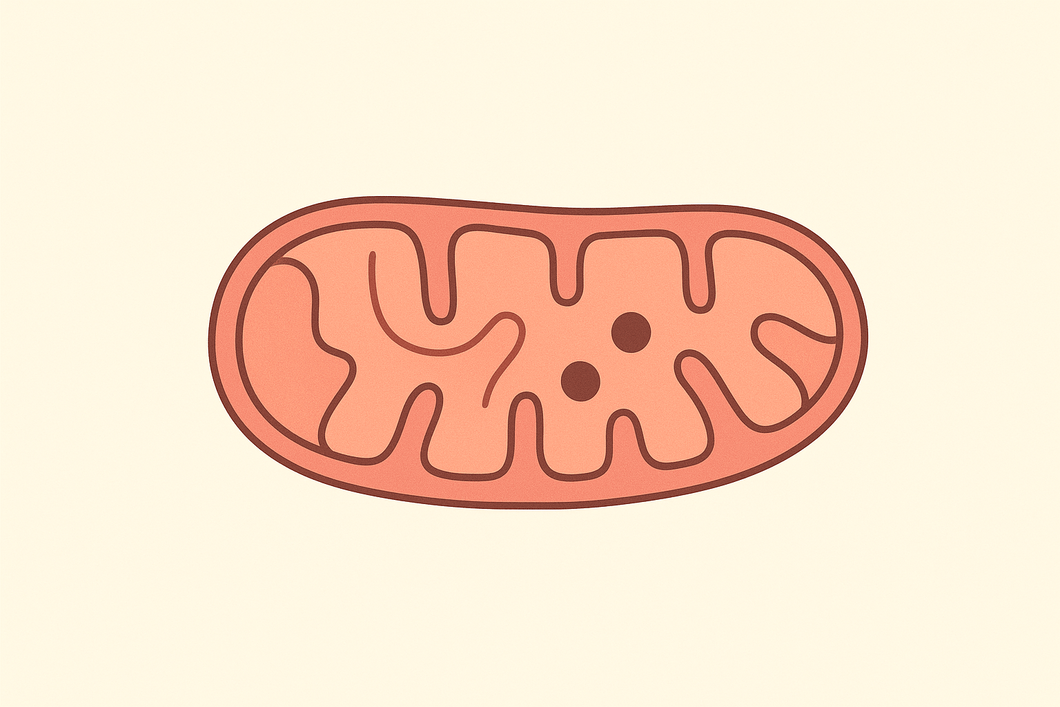
Structure of a Mitochondrion
Every mitochondrion has a four-layer architecture. Each layer has a distinct job.
Outer Membrane
The outer mitochondrial membrane is a smooth phospholipid bilayer freely permeable to small molecules (under about 5,000 daltons) through porin channels. It defines the outer boundary of the organelle and separates it from the cytosol. It does not participate directly in energy production.
Intermembrane Space
Between the outer and inner membranes sits a narrow gap, the intermembrane space. Protons pumped during the electron transport chain accumulate here, building the gradient that drives ATP synthase. The proton concentration in the intermembrane space is roughly 10x higher than in the matrix.
Inner Membrane and Cristae
The inner membrane is folded into convolutions called cristae. The folding multiplies the surface area many times over. This is where the electron transport chain (complexes I through IV) and ATP synthase (complex V) sit, embedded in the membrane. The inner membrane is highly impermeable, which is what makes the proton gradient possible. Only ATP synthase and specific transporters can let protons cross.
Matrix
The matrix is the innermost compartment. It contains the enzymes of the Krebs cycle (citric acid cycle), the enzymes of fatty acid β-oxidation, mitochondrial ribosomes, and multiple copies of mitochondrial DNA. Mitochondrial ribosomes are smaller than eukaryotic cytoplasmic ribosomes and structurally closer to bacterial ribosomes, which is a major piece of evidence for the endosymbiotic origin.
How Mitochondria Produce ATP
ATP production from glucose happens in three sequential stages, and only the last two take place inside the mitochondrion.
- Glycolysis (cytosol). Glucose is broken into two molecules of pyruvate, producing 2 ATP and 2 NADH. Glycolysis happens in the cytosol, outside the mitochondrion. The pyruvate then enters the mitochondrial matrix.
- Krebs cycle (matrix). Each pyruvate is converted to acetyl-CoA, which enters the Krebs cycle. Per glucose, the cycle produces 6 NADH, 2 FADH₂, 2 GTP/ATP, and releases 6 CO₂ molecules. The CO₂ exhaled in respiration comes from the Krebs cycle running in the mitochondrial matrix.
- Oxidative phosphorylation (inner membrane). NADH and FADH₂ deliver electrons to the electron transport chain. As electrons hop through complexes I → III → IV, protons are pumped from the matrix into the intermembrane space. The resulting proton gradient drives ATP synthase, which produces ATP. Oxygen is the final electron acceptor at complex IV, reduced to water. This is why aerobic respiration requires oxygen.
The bottom line: aerobic respiration yields roughly 30-32 ATP per glucose molecule, of which ~28 come from oxidative phosphorylation inside the mitochondrion. Anaerobic respiration (without mitochondrial involvement) yields just 2. This 15x improvement is what makes large multicellular life possible.
Mitochondrial DNA
Mitochondria contain their own DNA, separate from the nuclear genome. In humans, mitochondrial DNA (mtDNA) is a small circular molecule of 16,569 base pairs encoding 37 genes: 13 protein-coding genes (all components of the electron transport chain), 22 transfer RNAs, and 2 ribosomal RNAs. Every mitochondrion contains 2 to 10 copies of mtDNA. Every cell contains hundreds to thousands of mitochondria. So a single cell may carry tens of thousands of copies of mtDNA.
Three properties of mitochondrial DNA matter:
- Maternal inheritance. mtDNA is inherited almost exclusively from the mother. The sperm’s few mitochondria are tagged with ubiquitin at fertilization and destroyed. This makes mtDNA a clean tool for tracing maternal lineages, including in deep population genetics.
- High mutation rate. mtDNA accumulates mutations roughly 10-17 times faster than nuclear DNA, because the mitochondrial matrix is a chemically hostile environment (reactive oxygen species are produced as ETC byproducts) and mtDNA has limited repair machinery.
- Heteroplasmy. Because a cell carries many mtDNA copies, mutations can be present in only some copies. Symptoms of mitochondrial disease often depend on what fraction of the cell’s mtDNA carries the mutation.
Endosymbiotic Theory
Mitochondria were almost certainly free-living alpha-proteobacteria about 1.5 to 2 billion years ago. A primordial archaea-like host cell engulfed one but did not digest it. The bacterium kept its own DNA and divided independently inside the host, exchanging energy for protection and nutrients. Over time, most of the bacterium’s genes migrated to the host nucleus, leaving the modern stripped-down 16,569 bp mtDNA.
The endosymbiotic theory is supported by multiple independent lines of evidence:
- Mitochondria have their own double membrane (inner from the original bacterium, outer from the host’s engulfing vesicle).
- Mitochondrial DNA is circular, like bacterial DNA, and not associated with histones.
- Mitochondrial ribosomes are 70S (closer to bacterial 70S than eukaryotic 80S).
- Mitochondria divide by binary fission, not by mitosis.
- Antibiotics that target bacterial ribosomes (e.g., chloramphenicol) also affect mitochondrial ribosomes.
Mitochondrial Diseases
Mitochondrial dysfunction underlies a growing number of human diseases. Because mitochondria are present in essentially every tissue and energy demand varies wildly by tissue, mitochondrial diseases hit the most energy-demanding organs hardest: brain, heart, skeletal muscle, eyes, ears.
- Leber’s hereditary optic neuropathy (LHON) causes sudden vision loss in young adults due to mtDNA mutations affecting Complex I.
- MELAS (mitochondrial encephalopathy, lactic acidosis, and stroke-like episodes) is caused by a specific tRNA mutation.
- Kearns-Sayre syndrome involves large mtDNA deletions and causes muscular and cardiac symptoms.
- Parkinson’s, Alzheimer’s, and Type 2 diabetes all show mitochondrial dysfunction as a major component, though not always as the primary cause.
Mitochondrial replacement therapy (sometimes called three-parent IVF) was approved in the UK in 2015 to allow women with mitochondrial disease to have biological children without passing the mutation on. The technique transfers nuclear DNA from the mother into a donor egg with healthy mitochondria.
Related study notes: Protein, Enzyme, Nucleic Acid.
Frequently Asked Questions
Why are mitochondria called the powerhouse of the cell?
Because they generate about 90% of the ATP a typical cell uses. ATP is the molecular energy currency that drives nearly every active process in the cell. Aerobic respiration in mitochondria produces roughly 30-32 ATP per glucose, compared with only 2 ATP from anaerobic glycolysis alone.
What is the function of the inner mitochondrial membrane?
The inner mitochondrial membrane is folded into cristae and holds the electron transport chain (Complexes I to IV) and ATP synthase (Complex V). The membrane is highly impermeable to protons, which lets the cell build a proton gradient between the intermembrane space and the matrix. ATP synthase uses that gradient to make ATP.
Do mitochondria really have their own DNA?
Yes. Human mitochondrial DNA is a circular molecule of 16,569 base pairs that encodes 37 genes essential for the electron transport chain. mtDNA is inherited almost entirely from the mother, mutates about 10-17 times faster than nuclear DNA, and is a key tool for tracing maternal lineages in population genetics.
What is the endosymbiotic theory?
The endosymbiotic theory says that mitochondria originated as free-living alpha-proteobacteria that were engulfed by an early eukaryotic ancestor cell roughly 1.5 to 2 billion years ago. Instead of being digested, the bacterium survived inside and over time evolved into the organelle we now call the mitochondrion. The double membrane, circular DNA, bacterial-style ribosomes, and binary fission are all evidence.
How many mitochondria does a cell have?
It varies wildly by cell type. A red blood cell has zero (it loses them during maturation to maximize hemoglobin space). A typical liver cell has 1,000 to 2,000. A heart muscle cell has 5,000 to 8,000 (about 30% of cell volume). High-energy-demand cells have many more mitochondria than low-demand cells.
Why are mitochondrial diseases often passed from mother only?
Because mitochondrial DNA is inherited from the mother. At fertilization, the sperm’s mitochondria are tagged with ubiquitin and destroyed; only the egg’s mitochondria are passed on to the zygote. Any mtDNA mutation a mother carries is transmitted to all her children, but a father’s mtDNA mutations are not passed to his offspring.